肝臟與膽道
考前可由頭掃到底,建立「先定位、再看血流、再看顯影、最後把臨床背景放回去」的肝膽判讀流程。
目的:考前可由頭掃到底,建立「先定位、再看血流、再看顯影、最後把臨床背景放回去」的肝膽判讀流程。本文刻意不依年份、題號或單題順序編排;所有題源足跡與去留理由見 本章題源稽核。
一、章首判讀地圖:拿到肝膽影像時先做什麼
1. 先回答五個總問題
- 病變是 diffuse 還是 focal?
- diffuse:脂肪、鐵沉積、纖維化/肝硬化、壅血、急性肝炎、瀰漫浸潤。
- focal:先確認是真正 mass,還是 focal fat、灌流差、血管或膽管造成的 pseudolesion。
- 病人是否屬 HCC 高風險族群?
- 肝硬化、慢性 HBV 等適用族群,才進入 LI-RADS 的診斷語境。
- 非高風險肝臟即使有 arterial hyperenhancement,也不能直接套 LR-5。
- 病灶的血流與動態顯影屬哪一型?
- nonrim APHE、washout、capsule:偏 hepatocellular pathway。
- rim APHE、targetoid appearance、progressive delayed central enhancement:偏非 HCC 惡性病灶。
- peripheral discontinuous nodular enhancement、向心填充:先想 hemangioma。
- 病灶有沒有細胞/組成線索?
- fat、blood product、iron、fibrosis、mucin、necrosis、bile、restricted diffusion、hepatocyte uptake。
- 有沒有改變處置的併發症?
- vascular invasion、active bleeding、rupture、biliary obstruction、abscess、infarction、transplant vascular compromise、post-treatment viable tumor。
2. 肝內 focal lesion 的固定閱讀順序
Step A:先看未增強影像
- CT:脂肪、出血、高密度蛋白液、鈣化、lipiodol。
- MRI:T1/T2、in-/opposed-phase、DWI/ADC、T2*/susceptibility、subtraction。
- US:囊實性、後方增強/陰影、血流、是否與膽管或血管相通。
Step B:看 arterial phase 的「型態」,不只看亮不亮
- nonrim homogeneous/heterogeneous APHE:HCC、FNH、HCA、hypervascular metastasis、shunt。
- rim APHE:iCCA、metastasis、combined tumor、abscess等,屬 LR-M 思路。
- peripheral discontinuous nodules:hemangioma。
- wedge-shaped、沿門脈分布、無 mass effect:灌流異常/arterioportal shunt。
Step C:portal venous 與 delayed phase 分開判斷
- 真正 washout 是病灶相對肝實質變暗,且需注意是否只是背景肝逐漸增強造成的 pseudowashout。
- progressive delayed enhancement 常代表 fibrous stroma;典型於 iCCA,也可見 scar、sclerosed lesion。
- capsule 要看是否為平滑、周邊、延遲增強;不把單純壓迫性 pseudocapsule 或鄰近纖維化誤算。
Step D:若用 gadoxetate(EOB)再看 hepatobiliary phase
- HBP iso/hyperintensity 代表存在 OATP transporter 或保留 hepatocyte function,最常幫助 FNH,但並非良性專屬。
- HBP hypointensity 是「缺乏正常 hepatocyte uptake」的敏感線索,可支持惡性,卻不具 HCC 特異性。
- gadoxetate 的 transitional phase 容易因正常肝實質快速增強造成 pseudowashout;LI-RADS 的 nonperipheral washout 應在 portal venous phase 評估。
Step E:最後才把臨床背景放回來
- 年齡、性別、慢性肝病、原發癌、口服避孕藥/anabolic steroid、發燒、免疫抑制、外傷、移植、介入史。
- 影像型態優先;臨床背景是調整 prior probability,不是替影像硬套診斷。
二、解剖與 hemodynamics:看懂 segment、灌流與假病灶
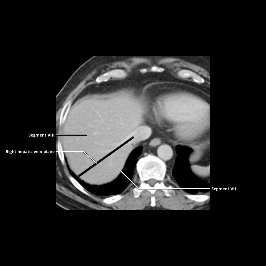
圖 S03-FIG-01|CECT/axial:上腹部軸位 CECT:右肝靜脈走在右葉內,作為 Couinaud segment VII/VIII 的分界;在 portal vein 平面以下同一靜脈面也分隔 V/VI。 來源:Liver
1. Couinaud 分段的核心
- Portal vein branches 位於 segment 中央,帶入 portal triad。
- Hepatic veins 位於 intersegmental plane,把肝臟切成 sectors。
- Middle hepatic vein 大致沿 Cantlie line,自 gallbladder fossa 指向 IVC,分隔右、左功能性肝葉。
- Right hepatic vein 分隔右前、右後 sector;left hepatic vein 分隔左內、左外 sector。
- Portal bifurcation 平面大致把上方 segment 7/8/4a/2 與下方 6/5/4b/3 分開。
- Caudate lobe(segment I) 可由左右 portal branches 供血,並有短 hepatic veins 直接回流 IVC,因此在 Budd–Chiari syndrome 與部分 cirrhosis 可相對肥大。
2. 常考的膽道與血管變異
- Right posterior sectoral duct 可能低位匯入 common hepatic duct、left hepatic duct,甚至 cystic duct;術前 MRCP 要主動找。
- Trifurcation、accessory hepatic duct 與 aberrant right hepatic artery 是 cholecystectomy/肝切除的重要風險。
- Replaced right hepatic artery 常來自 SMA;replaced left hepatic artery 常來自 left gastric artery。
- Cystic artery 多由 right hepatic artery 發出,但變異多;膽囊頸附近的 Calot triangle 是手術危險區。
3. 肝臟雙重供血與 THAD
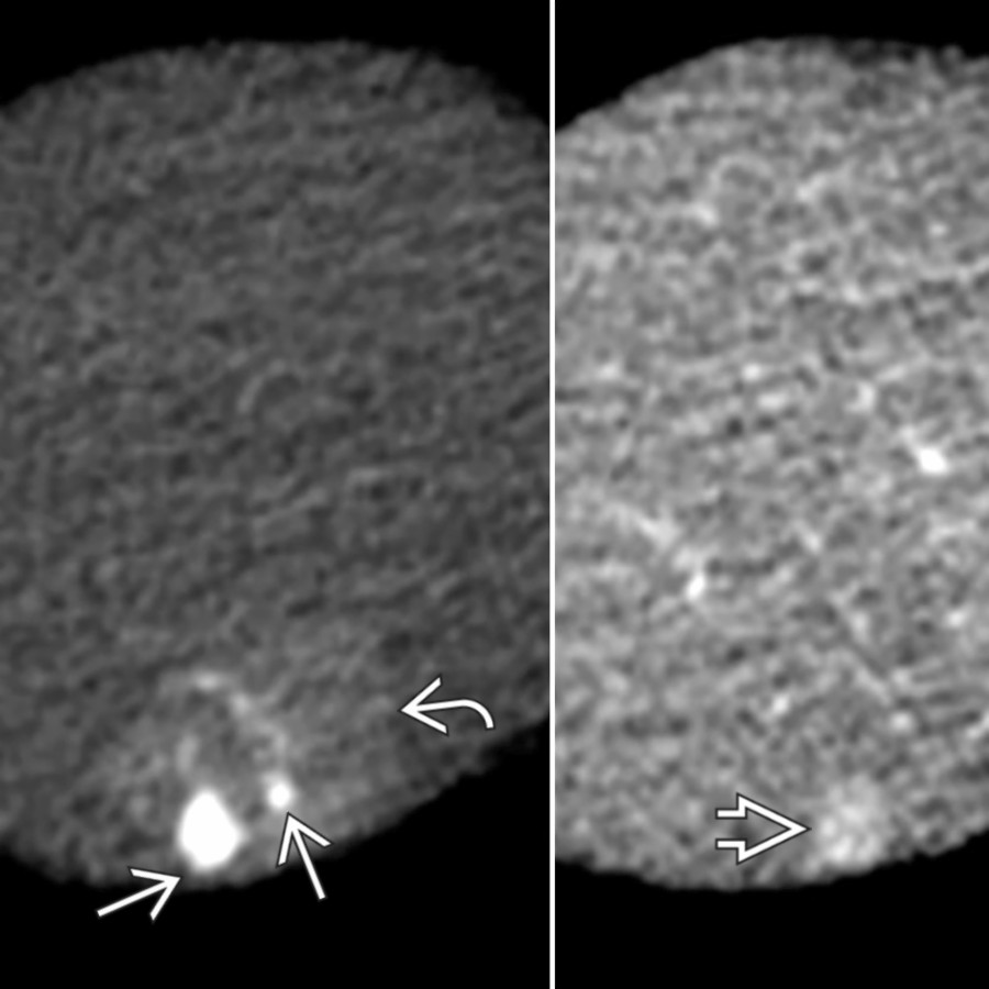
圖 S03-FIG-02|CECT, arterial phase/axial:動脈期 CECT:白實箭頭為肝頂部 flash-filling hemangiomas,白彎箭頭標示鄰近 THAD;後者是灌流改變,不應另算一顆腫塊。 來源:Hepatic Cavernous Hemangioma
正常肝實質約以 portal venous inflow 為主、hepatic arterial inflow 為輔。當局部 portal flow 降低或出現 arterioportal shunt,hepatic arterial buffer response 使該區 arterial phase 暫時變亮,形成 transient hepatic attenuation/intensity difference(THAD/THID)。
支持灌流異常而非腫瘤的線索:
- wedge-shaped 或 geographic,尖端朝 hepatic hilum;
- 沿 portal territory 分布;
- 血管穿越而無 displacement;
- 無真正 mass effect,T2/DWI 不形成對應實體;
- portal venous/delayed phase 回復等密度或等訊號。
常見原因包括 portal vein branch thrombosis、膽管炎/膽囊炎鄰近充血、arterioportal fistula、腫瘤壓迫門脈、介入後變化。THAD 是血流現象,不是診斷本身;一定要追查造成 portal flow 改變的原因。
4. Portal vein thrombosis:bland 還是 tumor in vein
| 線索 | Bland thrombus | Tumor in vein |
|---|---|---|
| 血栓內增強 | 無 | 有,最好用 subtraction 確認 |
| 血管徑 | 可正常或縮小 | 常擴張 |
| 與腫瘤連續 | 不一定 | 常直接連續 |
| DWI | 可限制,非特異 | 常與原腫瘤相似 |
| 其他 | cavernous transformation、誘因 | HCC 最典型,也見其他惡性腫瘤 |
- 慢性 portal vein occlusion 可形成 cavernous transformation:porta hepatis 多條蜿蜒 collateral vessels,並可能造成 portal biliopathy。
- 看到 portal vein 內「增強的軟組織」時,比單純 DWI restriction 更支持 tumor in vein。
5. Portal hypertension 與 collateral pathways
- 影像線索:splenomegaly、ascites、recanalized paraumbilical vein、gastroesophageal varices、splenorenal shunt、mesenteric congestion、portal flow reversal。
- Portosystemic collaterals 是壓力旁路;不要只看到靜脈曲張而漏掉原因,如 cirrhosis、portal vein thrombosis、Budd–Chiari syndrome。
- Clinically significant portal hypertension(CSPH)以 HVPG ≥10 mmHg 定義。 非侵入性 elastography 與 platelet count 可協助 rule-in/rule-out,但影像題通常先考 collateral、splenomegaly 與血流方向。[外部校正:Baveno VII]
6. Budd–Chiari syndrome、congestive hepatopathy 與 infarction
Budd–Chiari syndrome
- hepatic veins/IVC outflow obstruction;可見 hepatic vein 不顯影、狹窄或膜狀阻塞。
- acute:肝腫大、ascites、斑駁增強、周邊低灌流。
- chronic:caudate hypertrophy、肝形變、intrahepatic collaterals、再生結節。
- caudate 因直接流入 IVC 可相對保留或增強。
Congestive hepatopathy
- dilated IVC/hepatic veins、contrast reflux、periportal edema、heterogeneous “nutmeg” enhancement、右心衰竭徵象。
- 慢性可進展為 cardiac cirrhosis;arterial phase 的斑駁增強不可誤認多發腫瘤。
Hepatic infarction
- 肝有雙重供血,單一 portal 或 arterial occlusion 未必造成大範圍 infarction;移植肝、休克或合併雙重供血受損時風險升高。
- 通常是周邊 wedge-shaped nonenhancement,之後可能萎縮、液化或感染。
三、Dynamic CT/MRI、EOB 與 CEUS 的統一判讀框架
1. 常用 phase 與用途
| Phase | 主要用途 |
|---|---|
| Noncontrast | fat、blood、calcification、baseline density/signal |
| Late arterial | APHE、arterial anatomy、active bleeding |
| Portal venous | washout、portal vein、metastases、腹腔全貌 |
| Delayed/equilibrium | fibrous stroma、capsule、scar、bile leak 部分線索 |
| Transitional(gadoxetate) | 混合 extracellular 與 hepatocyte uptake;易出現 pseudowashout |
| Hepatobiliary phase | hepatocyte uptake、biliary excretion、small lesion detection |
2. 「Washout」的三個陷阱
- 沒有先證明 APHE:單純 delayed hypointensity 不是完整的 wash-in/washout pattern。
- 背景肝變亮造成 pseudowashout:尤其 gadoxetate transitional phase。
- 周邊 washout 判讀錯位:LI-RADS 要的是 nonperipheral washout;targetoid peripheral washout 是另一套惡性線索。
3. Subtraction、DWI 與 T2 的角色
- 出血、脂肪或蛋白質使病灶 precontrast T1 high,肉眼容易誤判增強;subtraction 是確認真增強的關鍵。
- DWI restriction 反映高細胞性、黏稠內容或缺血,不等同 malignancy:abscess、部分 benign lesion 也會限制。
- 很亮的 T2 “light-bulb bright” 支持典型 hemangioma/simple fluid;輕中度 T2 hyperintensity 比較廣泛。
- T2 dark 可見 fibrous tissue、iron、部分 fibrolamellar scar、sclerosed hemangioma。
4. EOB(gadoxetate)HBP:把它當 hepatocyte function map
| HBP 表現 | 常見解讀 |
|---|---|
| Iso/hyperintense | FNH 最典型;部分 β-catenin HCA、well-differentiated HCC 或保留 OATP 的病灶也可能出現 |
| Hypointense | 多數 metastases、iCCA、HCA、HCC;敏感但不特異 |
| Ring-like/targetoid hypointensity | 非 HCC 惡性病灶的重要輔助線索 |
重要觀念:
- FNH 的 uptake 來自 functioning hepatocytes 與異常 bile ductules;HCA 通常缺乏正常 bile ducts,故多數 HBP dark。
- HBP defect 可讓小 metastasis 或小 HCC 更顯眼,但最終分類仍要回到 dynamic pattern 與風險族群。
- 膽道排泄不佳可反映肝功能差或膽道阻塞;此時 HBP 品質下降,不能把低對比硬當病灶。
5. CEUS:真正的血池動態
- Microbubble 留在血管內,不像 CT/MRI extracellular agent 進入 interstitium;washout timing 與程度因此很重要。
- HCC 常見 arterial hyperenhancement + late、mild washout。
- iCCA/metastasis 等非 HCC 惡性病灶常見 early(約 60 秒內)或 marked washout。
- Hemangioma:peripheral discontinuous nodular enhancement、centripetal fill-in。
- FNH:快速 centrifugal/spoke-wheel filling,晚期多維持 iso-或 hyperenhancement。
- Abscess:周邊 septal enhancement、中央不增強;CEUS 可協助引流定位。
6. Technique-dependent detection traps:THI 與 partial volume
- 2026 新考點|Tissue harmonic imaging(THI):利用組織內產生的 harmonic frequency 成像,可減少 near-field clutter、reverberation 與 side-lobe artifact,使 liver/gallbladder boundary 與囊性病灶內部更乾淨;代價是深部 penetration 可能下降,肥胖或深部肝臟仍須回調 frequency、gain 與 focal zone,而不是固定全程開啟。
- 2026 新考點|Small-lesion partial volume:3 mm 低密度 metastasis 最容易被厚切面平均掉。真正有效的對策是取得/重建 thin sections,並縮小 reconstruction increment(可重疊重建);單純放大畫面不會恢復已在 acquisition/reconstruction 中遺失的 contrast。薄層提高 noise,需在 kernel、dose、迭代重建與檢出率間取捨。
這兩項改變的是病灶「能否被看見」,不是病灶生物學;確認技術品質後,仍須回到前述 enhancement、diffusion、T2 與 HBP 判讀軸。
四、瀰漫性肝病:先辨組成,再看形態與血流
1. Steatosis 與 focal fat
- CT:非增強肝密度下降;肝脾差值受 protocol、iron 與 contrast 影響。
- MRI chemical shift:opposed-phase 相對 in-phase 訊號下降最敏感。
- Focal fat/focal sparing 常在 gallbladder fossa、porta hepatis、segment IV、falciform ligament 周邊;呈 geographic、無 mass effect、血管正常穿越。
- Focal fat 可在 ultrasound 形成高回音假腫塊;MRI chemical shift 通常一槌定音。
- 2026 新考點|MRI-PDFF:應使用 confounder-corrected multiecho chemical-shift-encoded MRI,同步估算 PDFF 與 R2*,校正 T2* decay、fat spectral complexity 與其他干擾。Iron deposition 並不必然讓合格的 MRI-PDFF 產生『嚴重誤差』;真正限制是極重度鐵沉積造成訊號過快衰減、低 SNR 或模型失配。
2. Iron、copper 與其他沉積
- Iron 造成 T2/T2* shortening,影像越往長 TE 訊號掉得越多;GRE/R2* 可定量。
- Primary hemochromatosis 以 hepatocyte iron 為主,肝、胰可低訊號而脾相對保留;secondary hemosiderosis 常同時累及 reticuloendothelial system,包括脾與骨髓。
- Fat 與 iron 可互相干擾,應使用多回波 quantitative technique,而非只看單組 in-/opposed-phase。
3. Fibrosis 與 cirrhosis
形態序列
- early:右後葉與 segment IV 相對萎縮、caudate/左外葉相對肥大可逐漸出現。
- advanced:表面結節、裂隙增寬、肝門擴大、再生結節、portal hypertension。
- confluent hepatic fibrosis 常位於右葉前段或 medial segment,呈 wedge-shaped、capsular retraction、delayed enhancement;可模仿 infiltrative tumor,但通常沿既有纖維化分布且無 mass effect。
Elastography
- 測到的是 stiffness,不是純 fibrosis。急性發炎、膽汁鬱積、壅血、餐後狀態可使值升高。
- 判讀要同時看 technique、quality metrics 與臨床狀態;不要用單一 cutoff 脫離疾病背景。
4. Acute hepatitis 與 periportal edema
- 影像可正常,也可見 hepatomegaly、heterogeneous enhancement、periportal edema、gallbladder wall edema、ascites。
- Gallbladder wall thickening 在 systemic disease、hepatitis、heart failure、hypoalbuminemia 很常見;沒有局部壓痛、stone、distension 或周邊發炎時,不應直接診斷 acute cholecystitis。
5. Regenerative、dysplastic nodule 與 siderotic nodule
- Regenerative nodules 多無 arterialization,可能含 iron 而 T2*/SWI dark。
- Dysplastic nodule 尤其 high-grade 可逐步出現 arterial supply、HBP defect、diffusion restriction;尚未具典型 HCC major features 時常落在 LR-3/LR-4。
- Nodule-in-nodule、mosaic architecture、intralesional fat、corona enhancement 支持 hepatocarcinogenesis progression。
五、良性與感染性 focal lesions
1. Simple cyst、biliary hamartoma 與 peribiliary cyst
Simple cyst
- water attenuation、T1 dark/T2 bright、無增強、無 mural nodule;US 為 anechoic、thin wall、posterior enhancement。
- 出血或感染後可變複雜,但若有真正 enhancing mural nodule/septum,要改想 cystic neoplasm 或 metastasis。
2026 新考點|Biliary hamartoma
Biliary hamartomas(von Meyenburg complexes)
- 多發、細小、散在、T2 bright,通常不與 bile ducts 相通;可呈 “starry sky”。
- 與 Caroli disease 的差別:Caroli 是 segmental/saccular duct dilatation,與膽道相通且有 central dot sign。
Peribiliary cysts
- 沿 hilum/large portal tracts 成串,常見於 cirrhosis;本身不與 bile ducts 相通,但可壓迫膽管,容易誤認 duct dilatation。
2. Hemangioma
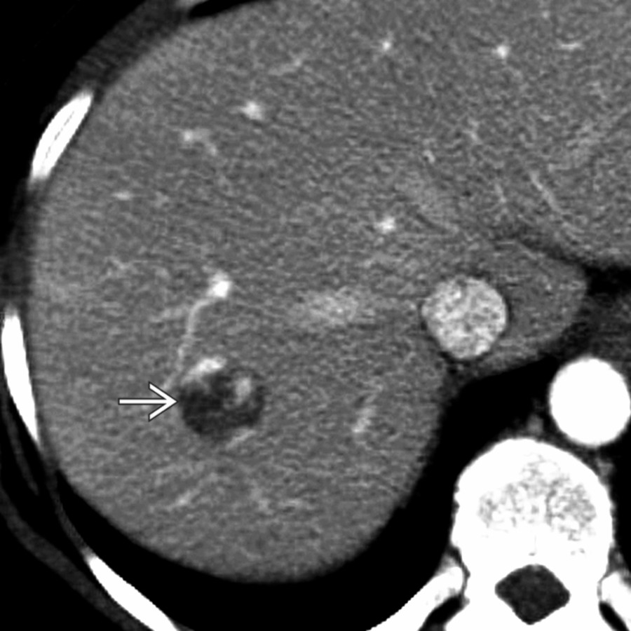
圖 S03-FIG-04A|CECT/axial:圖 1/2。門脈期 CECT 顯示周邊不連續結節狀增強,增強部分密度貼近血池;後續期相應觀察向心填充。 來源:Hepatic Cavernous Hemangioma
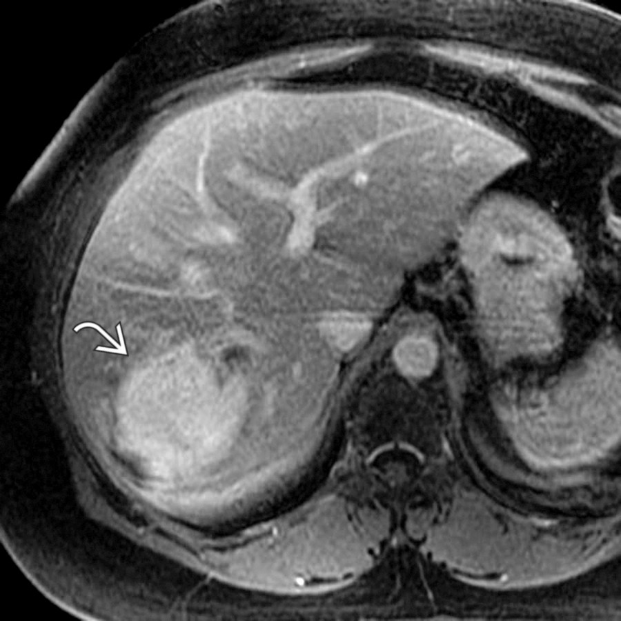
圖 S03-FIG-04B|T1 C+ FS MR/axial:圖 2/2。對比增強 T1 MRI 可見病灶由周邊向心填充,且增強程度接近血池,符合典型 hemangioma pattern。 來源:Hepatic Cavernous Hemangioma
典型 cavernous hemangioma:
- US homogeneous hyperechoic,無 halo;但 fatty liver 中可相對 hypoechoic。
- MRI very T2 bright。
- dynamic:peripheral discontinuous nodular enhancement,結節密度/訊號近 blood pool,逐步 centripetal fill-in。
變異與陷阱:
- small flash-filling hemangioma 可 arterial phase 均勻增強;看後續是否維持 blood-pool matching,而非真正 washout。
- giant hemangioma 可有 thrombosis、fibrosis、central scar,不一定完全填滿。
- sclerosed hemangioma 可縮小、capsular retraction、T2 不再極亮、延遲纖維化增強,模仿 malignancy。
- 典型 hemangioma 不需 biopsy;穿刺有出血風險且通常不能增加價值。
3. FNH 與 hepatocellular adenoma:先問「有沒有正常 bile duct/OATP」
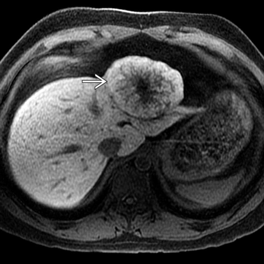
圖 S03-FIG-03A|Eovist-enhanced T1 MR, delayed phase/axial:圖 1/2。Eovist 注射後 20 分鐘 HBP:FNH 內對肝細胞特異性對比劑呈近均勻保留;中央瘢痕因無 hepatocytes 而不增強。 來源:Focal Nodular Hyperplasia
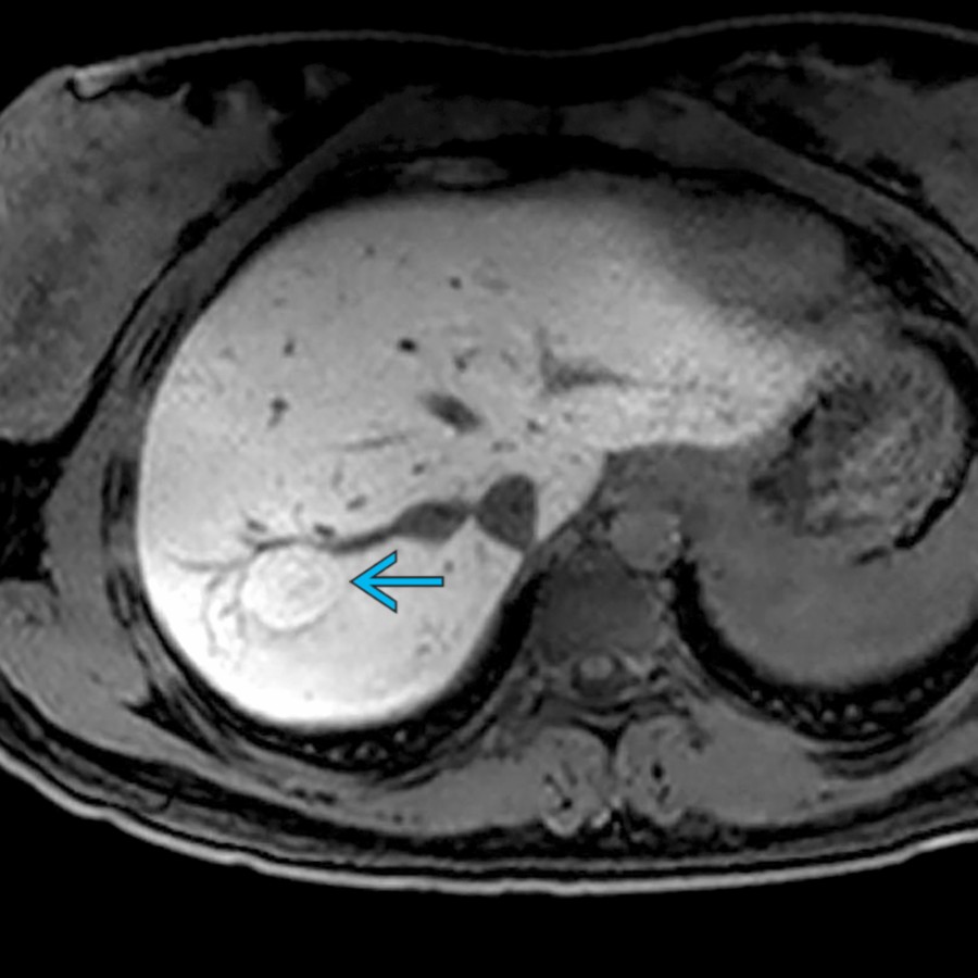
圖 S03-FIG-03B|Eovist-enhanced T1 MR, delayed phase/axial:圖 2/2。Eovist 20 分鐘 HBP:病灶相較背景肝仍呈輕度均勻高訊號(青箭頭),支持 FNH 的 OATP/hepatocyte uptake。 來源:Focal Nodular Hyperplasia
| 特徵 | FNH | HCA |
|---|---|---|
| 族群 | 常見年輕女性,非真正腫瘤性增生 | 女性、estrogen;男性/anabolic steroid;metabolic syndrome |
| 動脈期 | 強、相對均勻;可見 spoke-wheel | 強但常較 heterogeneous |
| Central scar | T2 bright、delayed enhancement | 通常無典型 scar |
| HBP | 多 iso/hyper | 多 hypo;β-catenin subtype 可 uptake |
| 出血/惡變 | 極低 | 有,依大小、性別與 subtype 而異 |
FNH
- central scar 並非每例可見;scar 的 T2 bright 與 delayed enhancement 有助和 fibrolamellar HCC 的 T2 dark、常鈣化 scar 區別。
- HBP uptake 是最實用的 discriminant,但不能單獨排除少數 uptake-positive HCA 或 HCC。
HCA subtypes
- HNF1A-inactivated:intralesional fat、opposed-phase signal drop;惡變風險較低。
- Inflammatory:T2 hyperintense、atoll sign、持續增強;出血較常見。
- β-catenin activated:可 HBP iso/hyper,malignant transformation risk 較高,男性尤其重要。
- 病灶內出血、脂肪、necrosis 使影像 heterogeneous;診斷與處置需結合性別、大小、成長與 subtype。
4. Pyogenic abscess、amebic abscess 與 microabscess
- Pyogenic abscess:cluster sign、double-target sign、周邊/septal enhancement、中央 diffusion restriction、gas;可能來自 biliary infection、portal spread、hematogenous spread 或術後。
- Necrotic tumor 也可限制 diffusion;發燒、周邊 edema、clustered small cavities 與臨床反應有助。
- Amebic abscess 常單發、右葉、周邊 edema;影像與 pyogenic abscess 重疊,需流行病史/serology。
- Candida microabscess:免疫抑制者多發小病灶,可見 bull’s-eye/wheel-within-wheel appearance,肝脾同時受累。
5. Hydatid disease 與其他寄生蟲
- Daughter cysts、detached membranes(water-lily sign)、wall calcification 是 hydatid cyst 的核心線索。
- 不同退化階段可由單純囊腫到複雜實性樣;未排除 hydatid 前不可隨意穿刺。
6. Peliosis 與 vascular benign lesions
- Peliosis hepatis 是多發含血腔隙,增強型態可變;與藥物、慢性病、惡性腫瘤相關。
- 影像若呈不典型多發 hypervascular lesion,尤其有 hemorrhage risk,需避免把所有血池樣病灶一概當 hemangioma。
六、HCC 與 LI-RADS:把分類當成可重複的決策樹
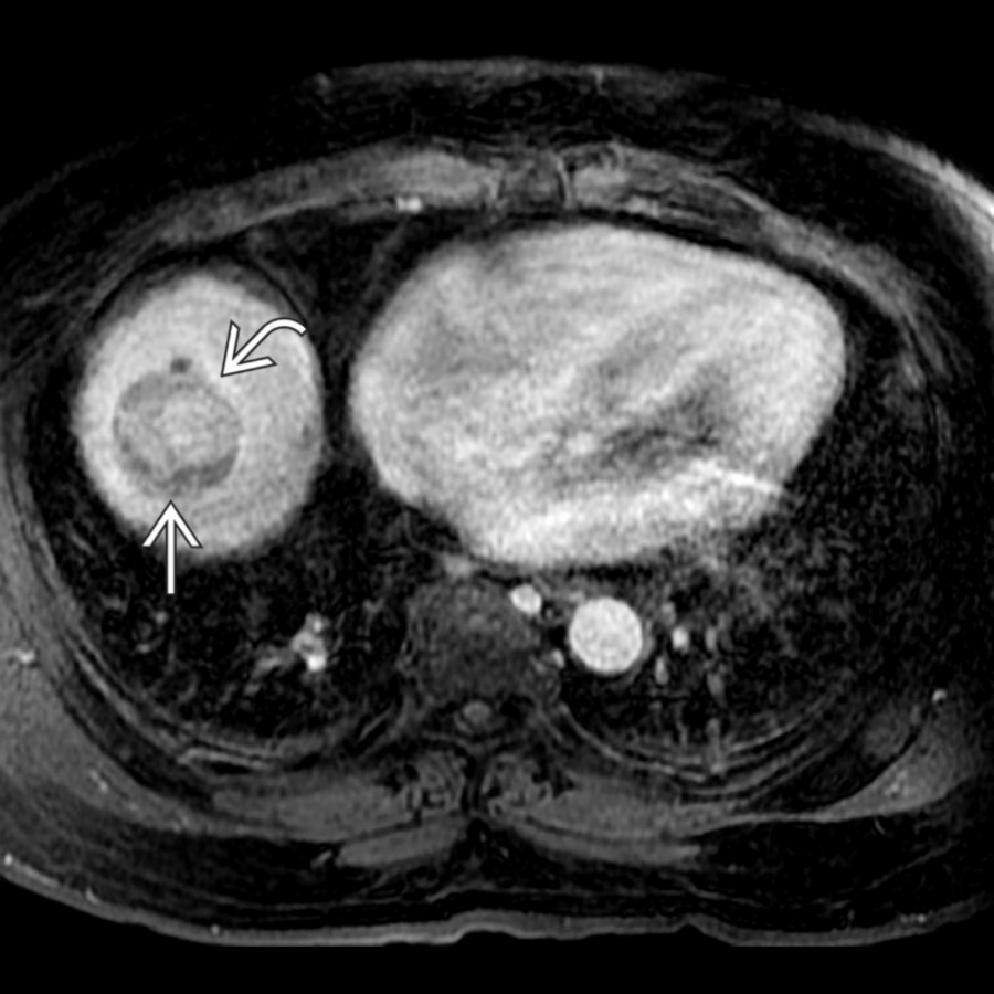
圖 S03-FIG-05A|T1 C+ FS MR/axial:圖 1/2。延遲期增強 T1 MRI:腫塊已 washout(白實箭頭),周邊可見 capsule(白彎箭頭),為 HCC 主要徵象。 來源:Hepatocellular Carcinoma
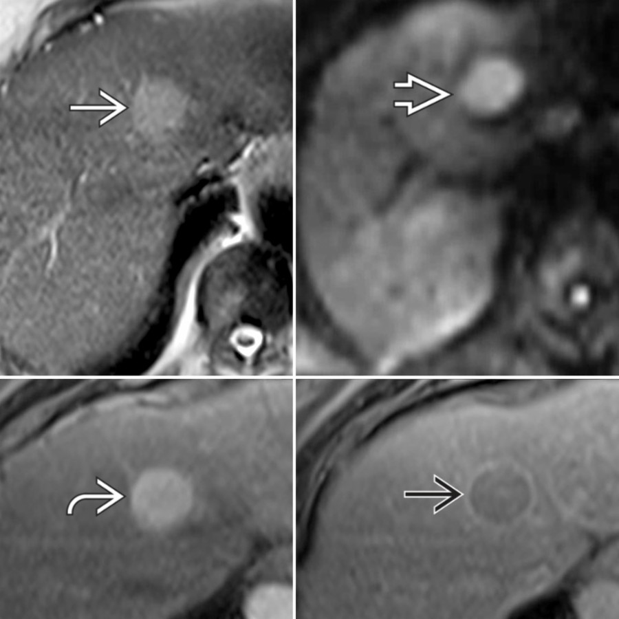
圖 S03-FIG-05B|MR/axial:圖 2/2。同一 HCC 的多序列 MRI:T2 高訊號、diffusion restriction、arterial hyperenhancement、靜脈期 washout 與 capsule 同時成立。 來源:Hepatocellular Carcinoma
1. 先確認 LI-RADS 是否適用
CT/MRI LI-RADS 用於具有 HCC 高風險的成人族群;不適用時應給 pattern-based differential,而不是硬貼 LR category。兒童、無高風險背景及部分先天性血管病變亦不宜直接套用。
2. Major features
- observation size
- nonrim arterial phase hyperenhancement(APHE)
- nonperipheral washout
- enhancing capsule
- threshold growth
版本校正重點(以題庫常用 CT/MRI LI-RADS v2018)
- threshold growth:6 個月內直徑增加 ≥50%。
- 10–19 mm observation:nonrim APHE + nonperipheral washout 可達 LR-5;只有 APHE + enhancing capsule 時仍不足 LR-5。
- 題目若引用較舊版的 12 個月成長窗口或已移除條件,要先辨識版本,不把舊敘述混入 v2018。[外部校正:ACR LI-RADS CT/MRI v2018 Core]
3. Category 的臨床直覺
| Category | 核心意思 |
|---|---|
| LR-1 | definite benign |
| LR-2 | probably benign |
| LR-3 | intermediate probability of malignancy |
| LR-4 | probably HCC |
| LR-5 | definite HCC by imaging criteria |
| LR-M | probably/definitely malignant,但不具 HCC 特異型態 |
| LR-TIV | unequivocal tumor in vein |
| LR-NC | technical limitation,無法分類 |
不要把 LR-M 當「不是 HCC」;它代表影像不能以高特異度指向 HCC,病理可仍是 atypical HCC、combined HCC-CCA 或其他 malignancy。
4. Ancillary features:可調整信心,但不能把 LR-4 升成 LR-5
Favoring malignancy in general
- restricted diffusion
- mild–moderate T2 hyperintensity
- HBP hypointensity
- corona enhancement
- fat sparing in a solid mass、iron sparing
- interval growth below threshold
Favoring HCC in particular
- intralesional fat
- mosaic architecture
- nodule-in-nodule
- nonenhancing capsule(依版本定義作 ancillary)
Ancillary features 可升/降一級,但 不能用來升到 LR-5;LR-5 必須靠 major-feature 規則。
5. LR-M:看到 targetoid 就先離開典型 HCC 路徑
常見 LR-M features:
- rim APHE
- peripheral washout
- delayed central enhancement
- targetoid diffusion restriction 或 HBP appearance
- infiltrative appearance、marked diffusion restriction、necrosis 等非特異惡性線索
主要 differential:iCCA、combined HCC-CCA、metastasis、atypical HCC。下一步取決於臨床情境,常需 biopsy 或 multidisciplinary discussion。
6. Tumor in vein
- 要求血管內有 unequivocal enhancing soft tissue;單純 thrombus 或鄰近 APHE 不夠。
- 可見 vein expansion、restricted diffusion、與原腫瘤連續。
- 影響 staging 與治療選擇,必須在報告中明確指出 vessel 與 extent。
7. HCC 的非典型與特殊型態
- Infiltrative HCC:界線不清、大片肝實質異常、常伴 portal vein tumor thrombus;APHE/washout 可不典型。
- Fibrolamellar HCC:年輕、通常無 cirrhosis;大、lobulated、central scar 常 T2 dark 且可 calcify,與 FNH 區分。
- Steatohepatitic/fat-containing HCC:intralesional fat 不代表良性。
- Spontaneously necrotic/hemorrhagic HCC:subtraction 幫助找殘存 viable nodule。
8. Treatment response:找「masslike viable tissue」,不是只看腔內是否還亮
- Viable:treated zone 內或邊緣有 nodular、masslike 或 thick irregular enhancement,呈 APHE、washout,或與治療前相似。
- Nonviable:無 masslike enhancement,或只有符合該療法的預期薄而平滑周邊增強。
- Equivocal:有可疑但不足以定性的 enhancement。
- Ablation 後立即可見 thin smooth rim、periablational hyperemia;殘存腫瘤較偏 eccentric nodular/irregular enhancement。
- TACE 後 CT 的 dense lipiodol 可遮蔽 enhancement;MRI subtraction 常較可靠。
- 評估 ablation margin 時,要把原腫瘤位置與 post-treatment defect 精準配準;單看病灶變小會太慢。
七、非 HCC 惡性病灶:以 targetoid、fibrosis、multiplicity 分流
1. Intrahepatic cholangiocarcinoma(iCCA)
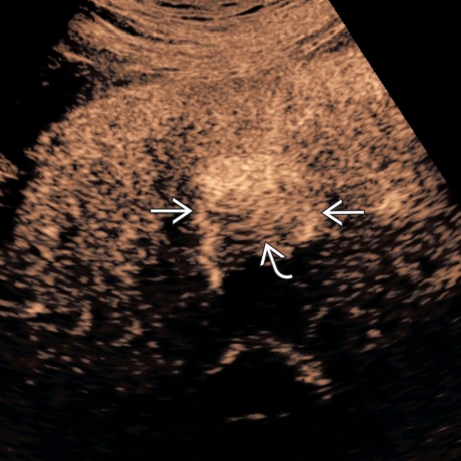
圖 S03-FIG-06A|contrast-enhanced ultrasound (CEUS)/transverse:圖 1/2。動脈期 CEUS:mass-forming iCCA 呈 rim APHE(白實箭頭)與中央低增強(白彎箭頭),走 LR-M/targetoid 路徑。 來源:Cholangiocarcinoma
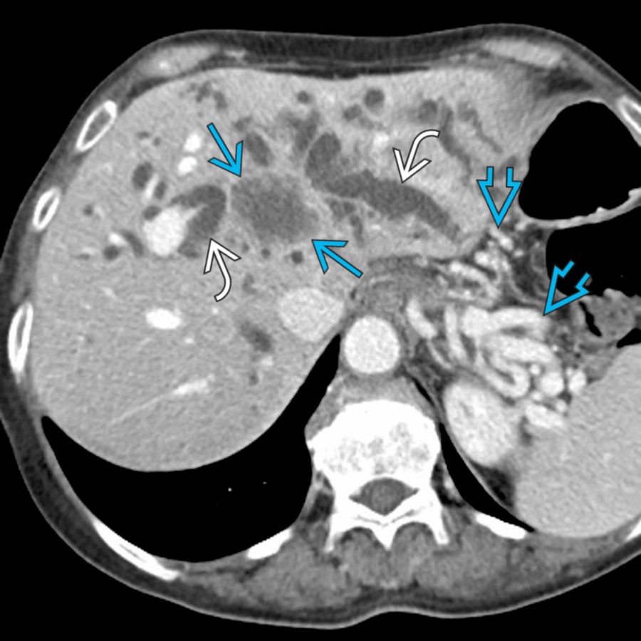
圖 S03-FIG-06B|CECT, portal venous phase/axial:圖 2/2。門脈期 CECT:肝門層級低密度 cholangiocarcinoma 侵犯肝實質,造成雙葉顯著肝內膽管擴張;另見門脈阻塞相關胃脾靜脈曲張。 來源:Cholangiocarcinoma
Mass-forming iCCA
- peripheral rim APHE,之後 progressive centripetal/delayed enhancement,反映 fibrous stroma。
- capsular retraction、distal biliary dilatation、satellite nodules、vascular encasement。
- DWI/HBP 可呈 targetoid appearance。
- 在高風險肝臟通常落入 LR-M,而非因「有 washout」就算 HCC。
Periductal infiltrating / intraductal growth
- periductal wall thickening、longitudinal spread、upstream ductal dilatation。
- intraductal tumor 可造成 duct expansion、mucin 與 papillary filling defects。
2. Combined HCC-CCA
- 同一腫瘤兼具 hepatocellular 與 biliary differentiation;影像可混合 APHE、washout、rim、targetoid、delayed fibrosis。
- 無法靠單一「像 HCC」的區塊排除 combined tumor;heterogeneity 與 LR-M features 應提高警覺。
3. Metastases
先用原發癌血流特性分類:
- hypovascular:colorectal、pancreatic、gastric 等,portal venous phase 較清楚。
- hypervascular:neuroendocrine tumor、RCC、melanoma、thyroid 等,arterial phase 必不可少。
- mucinous metastasis 可高 T2、calcification;necrotic metastasis 可呈 rim enhancement。
- 多發、不同大小、subcapsular 分布、target sign 支持 metastasis,但單發也不能排除。
- EOB HBP 對小 metastasis 敏感;DWI 可提高 detection,但 benign small cyst/hemangioma 仍需以 T2 與 enhancement 分辨。
4. Lymphoma
- 可單發、多發或 diffuse infiltration;常 homogeneous、restricted diffusion,enhancement 相對少。
- Vessel penetration without thrombosis(vessel floating)與 bulky adenopathy 可提示 lymphoma。
- 免疫抑制或 transplant 病人要想到 PTLD。
5. Vascular malignancies
Epithelioid hemangioendothelioma(EHE)
- 多發周邊結節,逐漸融合,capsular retraction。
- target appearance、lollipop sign(血管在病灶邊緣突然中止)具提示性。
Angiosarcoma
- 多灶、出血、壞死、progressive heterogeneous enhancement;可能引發 hemoperitoneum 或 consumptive coagulopathy。
- biopsy 有出血風險,需先做風險評估。
6. Cystic biliary neoplasms
- Mucinous cystic neoplasm of liver:多房、septum、mural nodule,通常不與膽道相通;多見女性,卵巢樣 stroma 是病理定義。
- IPNB:與膽管相通、intraductal papillary mass、duct expansion、mucin;可呈 segmental 或 diffuse ductal dilatation。
- 看到 enhancing mural nodule、thick septum 或 upstream ductal change,就不能以「complex cyst」結案。
八、膽管:先定阻塞層級,再分 stone、inflammation、tumor
1. 膽道擴張的固定算法
- 真的有擴張嗎? 考慮年齡、cholecystectomy 後狀態與肝功能。
- Intrahepatic、extrahepatic,還是兩者都有?
- transition point 在哪裡? hilar、mid-CBD、distal CBD/ampulla。
- 管腔內、管壁、還是外壓?
- lumen:stone、sludge、blood clot、parasite、intraductal tumor。
- wall:cholangitis、PSC、IgG4-related disease、CCA。
- outside:pancreatic head tumor、nodes、Mirizzi syndrome。
- 有沒有 cholangitis、abscess、atrophy 或 vascular involvement? 這些決定急迫性與可切除性。
2. Choledocholithiasis 與 MRCP pitfalls
- Stone 多為 T2 dark filling defect,可有 upstream dilatation。
- Mimics:air、flow artifact、blood clot、surgical clip、crossing vessel、sphincter contraction。
- Air 常位於 nondependent portion,並可見 susceptibility/CT gas;stone 多 dependent。
- 無 ductal dilatation 不排除小 stone;EUS 對微小 distal stone 很敏感。
3. Acute cholangitis
- 臨床核心是 infection + biliary obstruction;影像用於找阻塞原因與併發症。
- 影像可見 ductal wall thickening/enhancement、periductal edema、heterogeneous hepatic enhancement、abscess。
- 嚴重 cholangitis 需要 drainage;影像報告應說明阻塞 level、cause、可引流路徑與 portal/hepatic arterial anatomy。
4. PSC、IgG4-related sclerosing cholangitis 與 recurrent pyogenic cholangitis
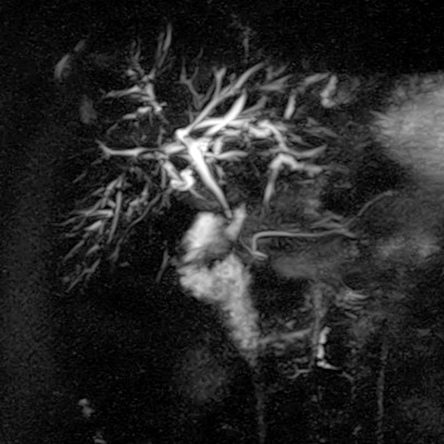
圖 S03-FIG-07|MRCP/coronal:冠狀位 MRCP MIP:肝內膽管多發短段狹窄與擴張形成 irregular beading;可視膽管分枝反而增多,反映擴張與狹窄並存。 來源:Primary Sclerosing Cholangitis
| 疾病 | 影像主軸 | 伴隨線索 |
|---|---|---|
| PSC | short multifocal strictures + intervening dilatation,beading;周邊 ducts pruning | IBD、CCA risk |
| IgG4-SC | long smooth strictures,常 distal CBD;可合併 hilar/intrahepatic | autoimmune pancreatitis、其他 IgG4 disease |
| Recurrent pyogenic cholangitis | intrahepatic pigment stones、central duct dilatation、pneumobilia、segmental atrophy | recurrent infection、東亞背景 |
- PSC 的 dominant/relevant stricture 若新出現不對稱增厚、mass、快速惡化或 CA19-9 上升,要排除 CCA。
- IgG4-SC 不應只靠 steroid response 事後診斷;先排除 malignancy。
5. Caroli disease、Caroli syndrome 與 choledochal cyst
- Caroli disease:先天性 nonobstructive saccular intrahepatic duct dilatation;central dot sign 是 portal radicle 位於擴張膽管內。
- Caroli syndrome:Caroli + congenital hepatic fibrosis,故同時有 portal hypertension。
- Todani choledochal cyst:
- I:extrahepatic fusiform/cystic dilatation;
- II:true diverticulum;
- III:choledochocele;
- IV:多發 intra-與/或 extrahepatic dilatation;
- V:Caroli disease。
- 需找 anomalous pancreaticobiliary junction、stone、cholangitis 與 malignancy complication。
6. Cholangiocarcinoma 的位置與手術語言
- Intrahepatic、perihilar、distal 三大類;同樣是 adenocarcinoma,影像與手術評估重點不同。
- Perihilar CCA:描述 longitudinal ductal extent(Bismuth–Corlette 僅反映 ductal distribution)、portal vein/hepatic artery involvement、lobar atrophy、future liver remnant、nodes 與 metastasis。
- Distal CCA:與 pancreatic head cancer、ampullary tumor 鑑別;看 lesion epicenter、pancreatic duct、duct wall thickening 與 vascular invasion。
- Ductal extent、vascular extent、lobar atrophy三者要分開寫,不能只報一個 Bismuth type。
7. Postoperative bile leak 與 biloma
- Biloma 通常為術床或 subhepatic fluid collection,可逐漸增大;若感染則壁增厚、gas、周邊發炎。
- 2026 新考點|Bile-leak MRI protocol:先在 gadoxetate 注射前完成 heavily T2-weighted MRCP,建立不受已排泄對比劑干擾的膽道解剖;再取得延遲 T1-weighted hepatobiliary images,直接尋找 contrast extravasation。
- Gadoxetate delayed hepatobiliary imaging 可直接顯示 contrast extravasation,但肝功能差時需更長延遲,陰性不能完全排除。
- ERCP、HIDA、percutaneous cholangiography 的選擇取決於疑似 leak level 與是否需同時治療。
九、膽囊:壁厚不是診斷,先找局部發炎與腔內病灶
1. Gallstone、sludge 與 tumefactive sludge
- Stone:US echogenic + posterior shadow,移動性有助;CT 可漏掉低密度 cholesterol stone。
- Sludge:low-level echoes、通常 layering、無內部血流。
- Tumefactive sludge 可呈 mass-like;無 Doppler/CEUS enhancement、改變姿勢或追蹤消失支持 sludge。若固定且有血流,需排除 neoplasm。
2. Acute cholecystitis
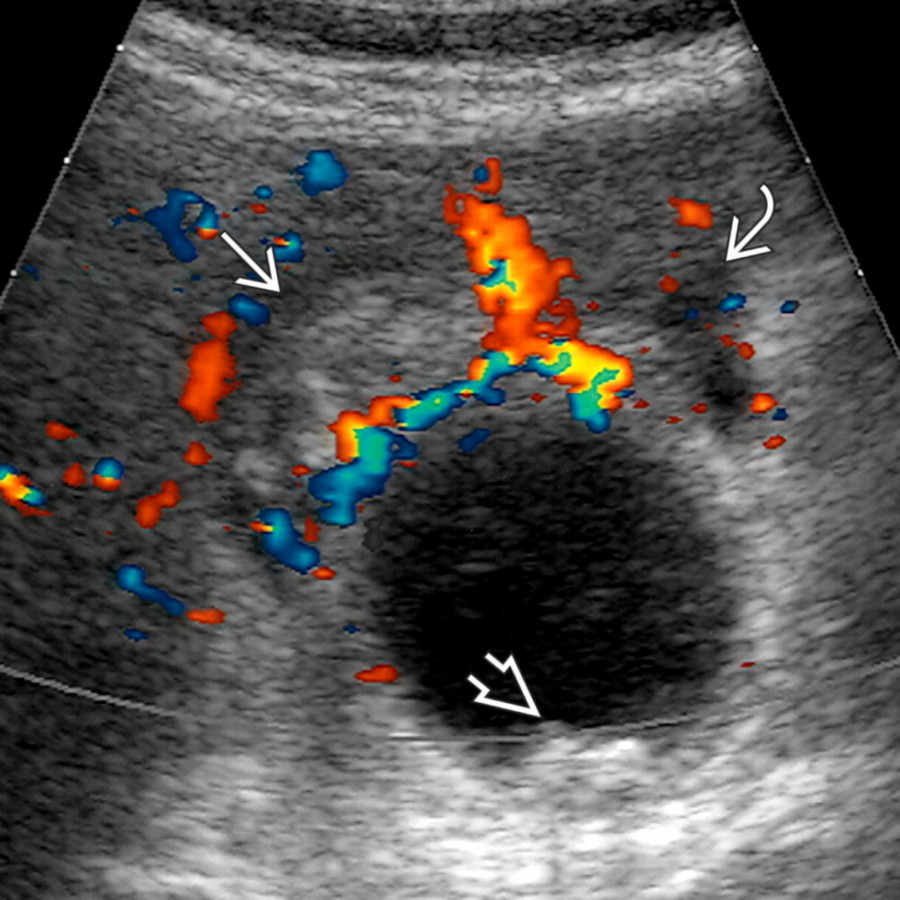
圖 S03-FIG-08|color Doppler ultrasound/transverse:橫切彩色 Doppler:壞疽性膽囊炎可見膽囊底周圍網膜脂肪 hyperemia、少量膽囊周圍液體與小結石。 來源:Acute Calculous Cholecystitis
影像線索要成組看:
- obstructing neck/cystic duct stone
- gallbladder distension
- wall thickening/stratification
- pericholecystic fluid、fat stranding
- hyperemia、sonographic Murphy sign
- adjacent hepatic hyperemia(segment IV/V THAD)
2026 新考點|TG18 組合架構
Tokyo Guidelines 的概念是:局部發炎徵象 + systemic inflammation 構成 suspected diagnosis,再加 characteristic imaging 才是 definite diagnosis。單獨 wall thickening 不夠。[外部校正:Tokyo Guidelines 2018]
Complications
- gangrenous:mucosal discontinuity、irregular wall、intraluminal membranes、decreased wall perfusion。
- perforation:wall defect、pericholecystic abscess、complex fluid。
- emphysematous:wall/lumen gas,常見 diabetic、elderly;CT 最敏感。
- hemorrhagic:high-density content/T1 high blood products,可能合併 hemobilia。
3. Chronic cholecystitis、XGC 與 carcinoma
- Chronic cholecystitis:shrunken gallbladder、stones、fibrotic wall;與 acute inflammation 疊加時仍可急性發作。
- Xanthogranulomatous cholecystitis(XGC):diffuse wall thickening、intramural hypoattenuating nodules、continuous mucosal line 可提示;但可侵入鄰近組織,與 carcinoma 高度重疊。
- Gallbladder carcinoma:mass replacing gallbladder、focal/asymmetric irregular wall thickening、polypoid mass、direct hepatic invasion、nodes/biliary obstruction。
4. Adenomyomatosis
- Rokitansky–Aschoff sinuses 是核心。
- US comet-tail/twinkling artifact;MRI “pearl necklace sign”。
- 可為 fundal、segmental 或 diffuse;segmental type 造成 hourglass configuration。
- 若外壁不規則、黏膜破壞、明顯 restricted diffusion 或 masslike invasion,不能只以 adenomyomatosis 解釋。
5. Gallbladder polyp
- Cholesterol polyp:多發、小、pedunculated、常有 comet-tail,無 shadow。
- Adenoma/neoplasm:單發、sessile、較大、內有 vascularity,伴 focal wall thickening 更可疑。
- 大小是處置的重要因素,但不是唯一因素;sessile morphology、快速成長、PSC、年齡與症狀會改變風險。考題常以約 10 mm 作傳統警戒門檻,臨床應依當地最新版 guideline 決策。
6. Mirizzi syndrome 與 gallstone ileus
- Mirizzi:impacted cystic duct/Hartmann pouch stone 外壓 common hepatic duct,造成 proximal biliary dilatation;長期可形成 cholecystocholedochal fistula。
- Gallstone ileus:mechanical SBO + ectopic gallstone + pneumobilia(Rigler triad);stone 常停在 terminal ileum,也可能造成 gastric outlet obstruction(Bouveret syndrome)。
十、急症、外傷、介入、移植與術後
1. Active bleeding、pseudoaneurysm 與 hemobilia
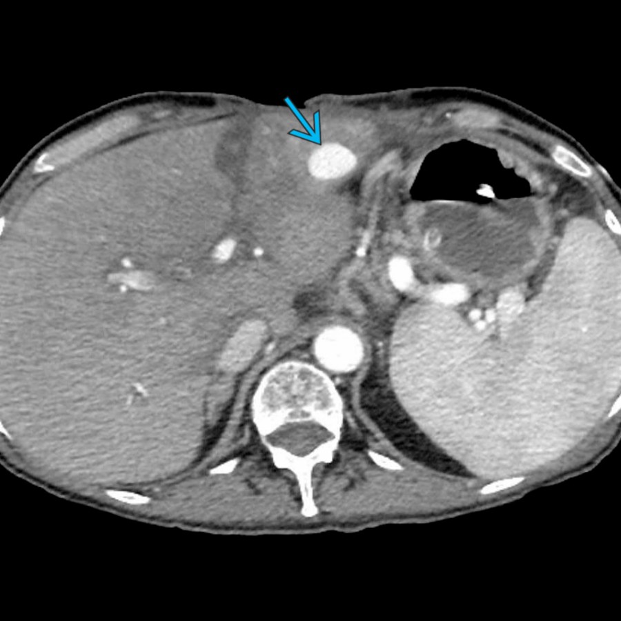
圖 S03-FIG-09A|CECT, arterial phase/axial:圖 1/2。動脈期 CECT:青箭頭標示左肝葉內 hepatic artery pseudoaneurysm,密度與同時期動脈血池一致。 來源:Hepatic Artery Pseudoaneurysm/Aneurysm
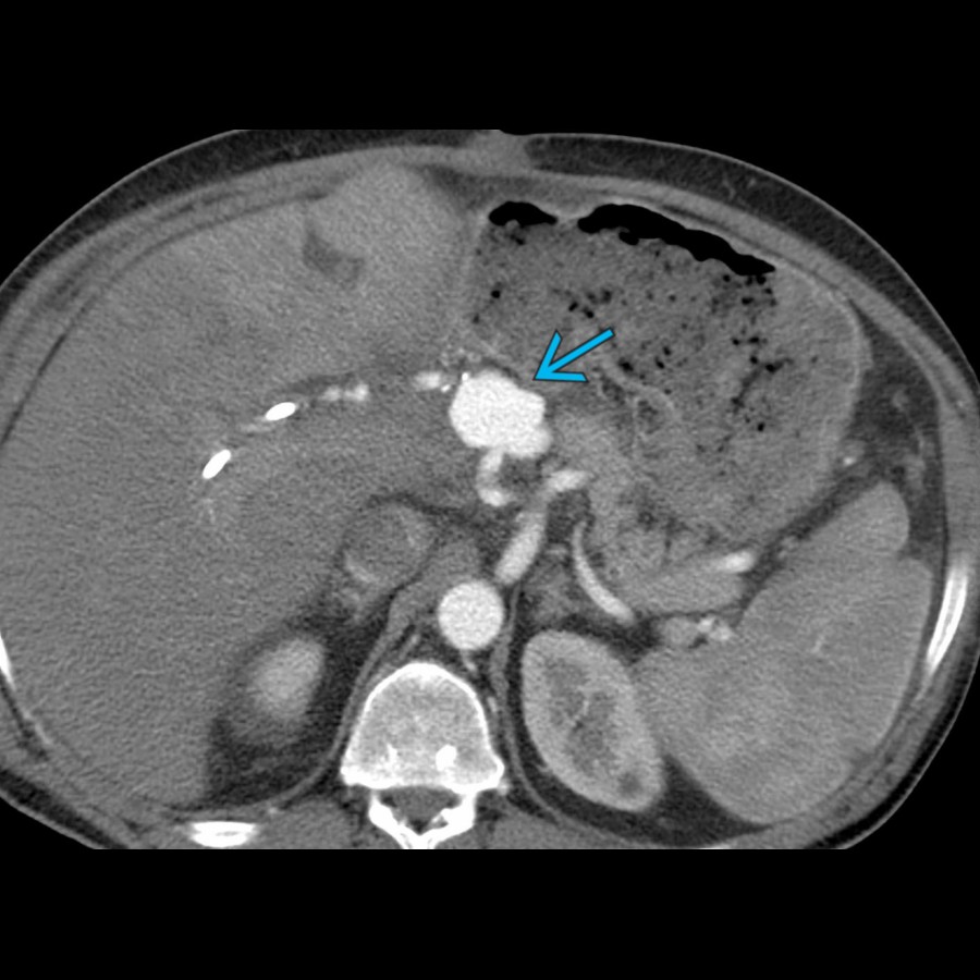
圖 S03-FIG-09B|CECT, arterial phase/axial:圖 2/2。動脈期 CECT:主 hepatic artery 在動脈吻合處呈局部瘤樣擴張(青箭頭),符合 anastomotic pseudoaneurysm。 來源:Hepatic Artery Pseudoaneurysm/Aneurysm
- Active extravasation:arterial phase 高密度,後續 phase 擴散/增大;要區分 pseudoaneurysm(局限、各期近血池)與單純高密度 clot。
- Hepatic artery pseudoaneurysm 可在 trauma、pancreatitis、surgery、biopsy、TACE 後出現;破裂可進腹腔、膽道或 GI tract。
- Hemobilia 的完整 Quincke triad(RUQ pain、jaundice、GI bleeding)並不常完整出現;影像找 high-density bile、arterial lesion、ductal clot。
2. Liver trauma:先報處置需要的資訊
- laceration depth、subcapsular/intraparenchymal hematoma、active bleeding、pseudoaneurysm、AV fistula、major venous injury、devitalized parenchyma。
- AAST grade 有助標準化,但臨床處置更取決於 hemodynamic stability、active arterial bleeding 與 associated injuries。
- Delayed complications:pseudoaneurysm、hemobilia、biloma、abscess、necrosis。
3. Spontaneous rupture
- HCC 與 HCA 是重要原因;影像找 subcapsular tumor、sentinel clot、capsular discontinuity、active extravasation。
- 先穩定與止血,常用 transarterial embolization;腫瘤 definitive treatment 另行規劃。
4. Ablation(RFA/MWA)
- 目標是完整覆蓋腫瘤並保留 safety margin;鄰近大血管有 heat-sink effect,MWA 較不受影響但仍需規劃。
- 鄰近 central bile duct、gallbladder、bowel、diaphragm 時需 hydrodissection、artificial ascites 或其他保護策略。
- Post-ablation gas、薄平滑 rim enhancement 可為預期;偏心 nodular enhancement 才像 residual viable tumor。
5. TACE/TAE 與 portal venous visualization
- Superselective catheterization 可提高 tumor dose 並減少正常肝傷害。
- Embolic endpoint 應依 technique、agent、tumor vascularity 與肝功能調整,不能簡化成固定「打到完全不流」。
- 在某些 HCC 的 superselective TACE,經腫瘤 sinusoid/peribiliary plexus 看見 portal venous branch opacification,可表示 embolic material 已穿過腫瘤微循環並達 portal side,常被視為充分填塞的線索;它不是 hepatic venous outflow,也不是所有個案都必須追求的普遍 endpoint。
- 非目標栓塞、arterioportal shunt、reflux 與肝功能儲備永遠優先於追求影像上的「漂亮終點」。
6. Portal vein embolization(PVE)與兩階段肝切除
- PVE 使預定切除側萎縮、future liver remnant(FLR)代償肥大。
- 報告要評估 portal anatomy、tumor progression、FLR volume/quality、是否有 portal thrombosis 或 nontarget embolization。
7. TIPS
- 目的:建立 portal vein 到 hepatic vein/IVC 的低阻力通道,降低 portal pressure。
- Doppler 評估 shunt patency、velocity trend、portal flow direction;單次絕對速度不如與 baseline 比較及整體 hemodynamics。
- Dysfunction 可由 stenosis、thrombosis、intimal hyperplasia 引起;同時評估 recurrent ascites/variceal bleeding 與 hepatic encephalopathy。
8. Liver transplant:血管先於膽道,膽道又依賴 hepatic artery
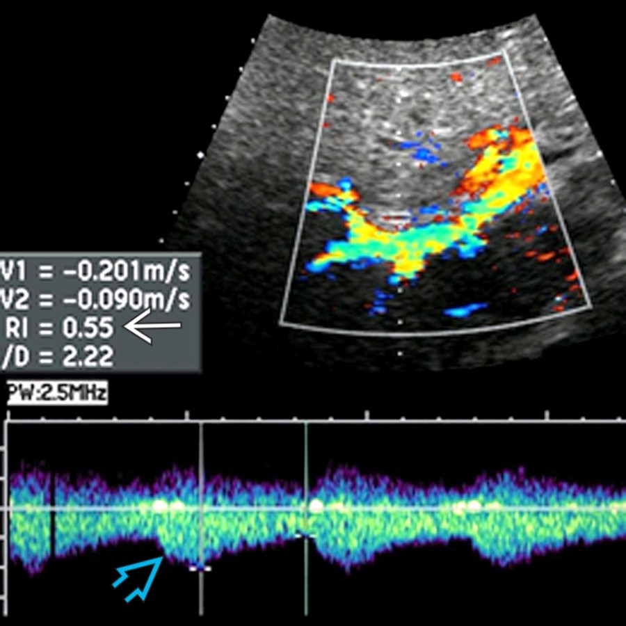
圖 S03-FIG-10|doppler ultrasound/spectral Doppler:右 hepatic artery spectral Doppler:收縮期上升遲鈍的 tardus-parvus 波形(青空心箭頭)合併低 RI(白實箭頭),提示上游狹窄。 來源:Liver Transplant Hepatic Artery Stenosis/Thrombosis
Hepatic artery
- Early HAT:無 flow、肝 infarction、rapid graft dysfunction;技術困難時 CTA/MRA/angiography 確認。
- Stenosis:tardus-parvus intrahepatic waveform、低 resistive index、延長 acceleration time;anastomotic focal high velocity。
- 肝內膽管主要依 hepatic arterial supply,因此 arterial compromise 可造成 ischemic cholangiopathy、biloma、nonanastomotic strictures。
Portal vein
- thrombosis、anastomotic stenosis、velocity step-up、portal hypertension signs。
Hepatic vein/IVC outflow
- waveform flattening 單獨不特異;要結合 focal narrowing、velocity gradient、graft congestion、ascites。
Biliary complications
- anastomotic stricture 多較短;nonanastomotic strictures 常與 ischemia、HAT、rejection 或 infection 相關。
- Bile leak、biloma、cast syndrome 亦常見。
Rejection
- 影像多不特異;排除 vascular/biliary obstruction 後仍需 clinical/laboratory/biopsy 判斷。
十一、兒童與先天性肝膽疾病
1. Neonatal cholestasis:biliary atresia vs neonatal hepatitis
支持 biliary atresia 的影像線索:
- triangular cord sign
- absent/small、形態異常 gallbladder,進食後不收縮
- nonvisualized common bile duct
- enlarged hepatic artery、subcapsular arterial flow
- 進展後 fibrosis、portal hypertension
沒有單一超音波徵象能完全排除;若臨床高度懷疑,需迅速完成 HIDA、biopsy 或 cholangiography 路徑,因 Kasai timing 影響預後。
2. Choledochal cyst 與 Caroli
- 兒童若有 jaundice、pain、mass 的 classic triad 實際並不常完整;US/MRCP 先確認囊性結構是否與膽道相通。
- Caroli syndrome 合併 congenital hepatic fibrosis,因此可同時有 recurrent cholangitis 與 portal hypertension。
3. Hepatoblastoma
- 多見幼兒,常為大型 heterogeneous mass,necrosis/hemorrhage/calcification,AFP 通常升高。
- 評估 PRETEXT sectors、hepatic/portal venous involvement、extrahepatic disease、肺轉移;重點不是只給診斷名稱,而是提供可切除性資訊。
4. Pediatric HCC
- 年齡較大,可與 chronic liver disease、metabolic disease 相關,但 fibrolamellar HCC 常無背景肝病。
- Conventional HCC 與 hepatoblastoma 的年齡、AFP、背景與影像重疊有限;仍需完整 staging。
5. Mesenchymal hamartoma
- 多見嬰幼兒,multicystic/stromal mass,可巨大;囊實比例依 myxoid stroma 與 bile ducts 而變。
- 與 undifferentiated embryonal sarcoma 的年齡、solid component、enhancement 與臨床行為區分;後者多見較大兒童且惡性。
6. Infantile hepatic hemangioma
- 可單發、多灶或 diffuse;hypervascular,可能造成 high-output heart failure、hypothyroidism。
- 大型 shunt lesion 可見 enlarged hepatic artery、tapered aorta below celiac axis、cardiomegaly;不能只把它當成人型 incidental hemangioma。
7. Congenital portosystemic shunt
- Abernethy malformation/congenital portosystemic shunt 可造成 hyperammonemia、pulmonary vascular complication,並伴 FNH-like nodules、HCA 或 HCC risk。
- 影像需說明 portal vein 是否存在、shunt anatomy、intrahepatic portal branches 發育與肝結節。
十二、最後速掃:考場決勝軸
A. 看 enhancement pattern
- Peripheral discontinuous nodules → centripetal fill-in:hemangioma。
- Homogeneous APHE + HBP uptake + central scar:FNH。
- APHE + fat/hemorrhage、HBP 多低:HCA;再看 subtype 與風險。
- Nonrim APHE + nonperipheral washout ± capsule(高風險肝):HCC pathway/LI-RADS。
- Rim APHE + targetoid + delayed central enhancement:iCCA/metastasis/combined tumor,走 LR-M。
- Wedge-shaped APHE、無 mass effect、後期消失:THAD/arterioportal shunt。
B. 看囊性病灶
- 無增強、water signal:simple cyst。
- daughter cyst/detached membrane:hydatid。
- cluster/double target + restriction:abscess。
- enhancing septa/mural nodule:MCN 或其他 cystic neoplasm。
- 與膽道相通、duct expansion/papillary tissue:IPNB/Caroli 等膽道病變。
C. 看膽道
- 先找 transition point,再分 lumen、wall、outside。
- Short multifocal strictures/beading:PSC。
- Long smooth stricture + autoimmune pancreatitis:IgG4-SC。
- Intrahepatic stones + central dilation + atrophy:recurrent pyogenic cholangitis。
- Saccular intrahepatic duct dilation + central dot:Caroli。
- Hilar stricture:除了 Bismuth type,還要報 vascular involvement、lobar atrophy 與 FLR。
D. 看膽囊壁
- wall thickening 是徵象,不是診斷。
- Stone + distension + local tenderness/hyperemia + pericholecystic inflammation:acute cholecystitis。
- Intramural cystic spaces/comet-tail:adenomyomatosis。
- Intramural nodules + preserved mucosal line:偏 XGC,但仍可能與 cancer 重疊。
- Focal irregular masslike thickening、mucosal disruption、liver invasion:gallbladder carcinoma。
E. 看治療後
- 薄、平滑、環狀增強可為預期反應。
- 厚、不規則、結節狀且呈原腫瘤 enhancement pattern:viable tumor。
- Lipiodol 遮蔽時用 MRI subtraction;不要只以 size shrinkage 判定成功。
F. 最容易犯的十個錯
- 非高風險肝臟也硬套 LI-RADS。
- 把 gadoxetate transitional-phase hypointensity 當 washout。
- 把 APHE wedge/THAD 當 HCC。
- 把 DWI restriction 當 malignancy 同義詞。
- 把 HBP uptake 當「一定良性」。
- 只因 gallbladder wall thickening 就診斷 cholecystitis。
- 看到 portal vein thrombus 卻不判斷 bland vs tumor。
- 把所有 rim-enhancing lesion 都叫 abscess,或把所有 targetoid lesion 都叫 metastasis。
- 治療後只看大小,不找 masslike enhancement。
- Perihilar CCA 只報 Bismuth type,漏掉 vascular extent、atrophy 與 FLR。
外部校正來源(只處理版本衝突與 guideline 缺口)
- American College of Radiology. CT/MRI LI-RADS v2018 Core 與官方 LI-RADS 資源頁:用於 threshold growth、10–19 mm LR-5 條件與 washout phase 的版本校正。官方入口:https://www.acr.org/Clinical-Resources/Reporting-and-Data-Systems/LI-RADS
- de Franchis R, et al. Baveno VII – Renewing consensus in portal hypertension. Journal of Hepatology. 2022;76:959–974. DOI: https://doi.org/10.1016/j.jhep.2021.12.022
- Yokoe M, et al. Tokyo Guidelines 2018: diagnostic criteria and severity grading of acute cholecystitis. J Hepatobiliary Pancreat Sci. 2018;25:41–54. DOI: https://doi.org/10.1002/jhbp.515
題源足跡
本章已用 本章題源稽核 所列 2021–2026 候選反查;題號僅留在 audit,避免破壞正文的概念連續性。正文中的外部標記只用於版本校正,不代表另寫成文獻回顧。